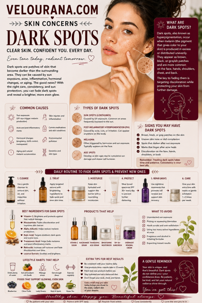
CATEGORY: Dark Spots
A dark spot is never just a dark spot.
The same biological event can leave a faint beige mark on one complexion, a deep brown patch on another, and a grey or blue-toned area on a third. Two people may experience the same degree of acne inflammation, use similar skincare and spend comparable time outdoors, yet the pigmentation that remains afterward can look dramatically different.
These differences are not cosmetic accidents. They reflect the way melanin is produced, packaged, transferred and distributed within different skin tones, as well as the depth at which pigment is located and the intensity of the trigger that created it.
Understanding these variations matters because pigmentation is often treated according to appearance alone. A person sees a dark area and assumes that all dark areas should respond to the same product. But colour provides only a partial clue. The biology underneath can vary considerably.
A superficial brown mark created by recent inflammation may behave very differently from a deeper grey-brown area. Melasma in medium or deeper skin may require a different strategy from a sun-induced lentigo in fair skin. A pigmentary change that appears subtle in one complexion may become highly visible in another because of contrast.
The relationship between skin tone and dark spots is therefore not simply about whether pigmentation is lighter or darker.
It is about how the pigmentary system responds, how easily inflammation stimulates melanin, how visible changes become against the surrounding complexion, and how carefully treatment must be balanced against the risk of creating more pigmentation.
That complexity explains why effective pigmentation care should never rely on one universal formula.
The Number of Melanocytes Is Not the Main Difference Between Skin Tones
One of the most common misconceptions about skin colour is that darker skin simply contains more melanocytes.
The reality is more nuanced.
People across a wide range of skin tones generally have a broadly similar number of melanocytes. The differences in visible colour are influenced much more by how actively those cells produce melanin, the type of melanin produced, and the size, distribution and persistence of melanosomes within surrounding skin cells.
Melanin exists in more than one form.
Eumelanin is brown to black and provides stronger photoprotective effects. Pheomelanin is red to yellow and behaves differently in response to ultraviolet radiation.
The relative amount and organisation of these pigments contribute to visible complexion.
In deeper skin tones, melanosomes tend to be larger, more heavily pigmented and distributed more individually within keratinocytes. In lighter skin, they are generally smaller and more frequently clustered.
These structural differences influence how the skin reacts after inflammation.
When melanocytes receive signals to increase pigment production, the resulting visual change may become considerably more pronounced in a complexion where pigment production is already highly active.
This is why post-inflammatory hyperpigmentation can become such a dominant concern in medium and deep skin tones.
The inflammation itself may resolve relatively quickly.
The pigmentary response can remain long afterward.
Contrast Determines How Noticeable Pigmentation Becomes
A dark spot does not exist visually in isolation.
We perceive it in relation to the surrounding skin.
A small increase in pigmentation can look extremely noticeable if the contrast between the mark and surrounding complexion is strong. Conversely, a considerable amount of pigment may appear less dramatic when it blends more closely with the baseline skin tone.
This creates an interesting paradox.
Fair skin can display highly obvious freckles, sun spots and post-inflammatory marks because brown pigment contrasts strongly against a pale background. Medium skin may show warm brown or grey-brown marks that linger after inflammation. Deep skin can develop pigmentation that appears very dark, grey, purple-brown or almost black depending on depth and lighting.
The visual experience is therefore influenced both by biology and optics.
Lighting further complicates perception.
Warm indoor lighting can make brown pigmentation appear softer. Cool daylight may emphasise grey or blue undertones. Strong side lighting can exaggerate texture and shadow, making a dark spot appear deeper than it actually is.
This is one reason judging pigmentation from photographs alone can be difficult.
A good clinical assessment considers not only colour but pattern, history, location and the events that preceded it.
Brown Pigment and Grey Pigment May Be Telling Different Stories
The colour of hyperpigmentation can offer clues about where pigment is located.
When melanin is concentrated mainly within the epidermis, pigmentation commonly appears brown.
When pigment extends deeper into the dermis, it may appear grey, slate, blue-grey or more muted.
This shift occurs partly because light interacts differently with pigment depending on how deeply it sits beneath the skin surface.
This distinction matters because depth influences how pigmentation responds to treatment.
Superficial pigment is more directly affected by the skin's normal turnover. As epidermal cells move toward the surface and are shed, excess melanin can gradually leave with them.
Deeper pigment behaves differently.
When melanin enters the dermis, cells known as melanophages can take up the pigment. Removal is considerably slower, which is one reason some marks persist long after the original inflammatory event has disappeared.
This can be especially relevant after severe inflammation.
A deeply inflamed acne lesion, aggressive procedure, burn or significant dermatitis may produce pigmentation that behaves very differently from a mild superficial blemish.
The assumption that a darker-looking mark simply requires a stronger exfoliating product therefore becomes problematic.
Depth cannot be scrubbed away.
Medium and Deep Skin Tones Often Pigment More Readily After Inflammation
Post-inflammatory hyperpigmentation is one of the most common pigmentary concerns in darker complexions.
The reason is not that inflammation itself is necessarily more severe.
The pigment-producing response can simply be more pronounced.
When inflammatory mediators stimulate melanocytes, the resulting increase in pigment may be substantial. A relatively minor injury can therefore leave a visible mark that remains long after the original problem has resolved.
Acne is a major example.
A person with deeper skin may find that the dark marks left after acne are more visually persistent than the active blemishes themselves. By the time one mark begins to fade, another breakout may have produced a new one.
This creates a layered pattern of pigmentation at different stages.
The oldest marks may appear faint.
More recent marks may appear richer brown.
Very recent lesions may still show inflammatory colour alongside developing pigment.
The complexion can therefore look uneven even when acne severity is relatively modest.
This is why acne management and pigmentation management should not be separated.
Preventing inflammation becomes one of the most effective ways to prevent future dark spots.
Redness Is Not the Universal Language of Irritation
Many skincare descriptions assume that irritated skin becomes visibly red.
That assumption works reasonably well in fair complexions, where vascular redness may be easy to detect.
In deeper skin tones, inflammation can be much less obviously red.
It may appear brown, purple, grey, darker than the surrounding skin or simply dull. Sometimes there is almost no visible colour change at all.
The person may instead notice burning, tightness, tenderness or increased sensitivity.
This matters enormously in pigmentation care.
If someone has been taught that redness is the main sign of irritation, they may continue using an overly aggressive routine because their skin "doesn't look irritated."
Yet the barrier may be compromised.
The pigmentation may worsen.
And the connection may be missed.
Sensation becomes important evidence.
If water suddenly stings, moisturiser burns, the skin feels tight after cleansing or previously comfortable products become uncomfortable, the routine may be too aggressive even without obvious redness.
In pigmentation-prone skin, ignoring these signs can create a cycle of inflammation followed by more pigment.
Fair Skin Has Different Pigmentary Vulnerabilities
Deeper skin tones often receive the most attention in discussions of post-inflammatory hyperpigmentation, but fair skin has its own characteristic pigmentary patterns.
Freckles may become more visible with ultraviolet exposure.
Solar lentigines often develop after years of accumulated sun exposure.
Post-inflammatory pigmentation can occur as well, particularly after acne or injury, although inflammatory marks in very fair skin may initially appear more red or pink because vascular changes can dominate before brown pigment becomes obvious.
This distinction is important.
A red mark after acne is not necessarily the same as post-inflammatory hyperpigmentation.
Post-inflammatory erythema involves persistent vascular redness rather than excess melanin. The two can coexist, but they respond differently.
A person may therefore spend months using pigment-targeting ingredients on what is primarily a vascular mark.
Again, diagnosis determines treatment.
Olive Skin Can Make Pigmentation Particularly Complex
Olive complexions often occupy an interesting middle ground.
They may tan readily, produce persistent post-inflammatory pigmentation and show melasma relatively clearly, yet visible redness can still appear depending on undertone.
Pigmentation may take on golden-brown, greenish-brown or grey-brown hues.
Because olive skin often tolerates sun without burning as quickly as very fair skin, there can also be a false sense of protection.
Tanning itself is evidence of a pigmentary response to ultraviolet exposure.
The absence of sunburn does not mean the skin is unaffected.
Repeated tanning contributes to cumulative photodamage and can worsen existing pigmentation.
This becomes particularly relevant when someone is trying to fade dark spots while intentionally maintaining a tan.
The two goals compete biologically.
The skin cannot be repeatedly encouraged to increase pigment production while also being asked to reduce uneven excess pigment as efficiently as possible.
Deeper Skin Requires Special Care With Procedures
Professional procedures can improve pigmentation, but the margin for error may be narrower in people who develop post-inflammatory hyperpigmentation easily.
Chemical peels, lasers, microneedling and other procedures intentionally create controlled injury or stimulation.
That controlled injury can be therapeutic.
It can also produce inflammation.
If the skin responds to inflammation with strong melanogenesis, inappropriate treatment settings or excessive intensity may result in worsening pigmentation.
This does not mean deeper skin should be excluded from procedures.
It means expertise matters.
The practitioner should understand how the treatment interacts with different skin types, how pigmentation risk changes with energy level or peel depth, and how to prepare and protect the skin before and after treatment.
The same principle applies to at-home devices.
A product marketed as universally safe may still produce very different outcomes across skin tones and individual sensitivities.
Pigmentation treatment should never treat skin colour as a cosmetic afterthought.
Melasma Can Look Different Across Complexions
Melasma commonly appears as symmetrical patches of hyperpigmentation on the face, but its colour can vary considerably.
On lighter skin it may look tan or light brown.
On medium skin it may appear deeper brown.
On deeper skin it may present as dark brown, grey-brown or mixed pigmentation depending on depth.
The pattern matters more than a single shade.
Melasma commonly affects the cheeks, forehead, upper lip and sometimes the jawline.
Hormonal influences, ultraviolet exposure, visible light and genetic predisposition can all contribute.
The condition is also notorious for recurrence.
This makes maintenance especially important.
Someone may achieve significant improvement and assume the pigmentation has been permanently eliminated. Then summer arrives, protection becomes inconsistent, and the patches gradually return.
This recurrence is not necessarily evidence that the original treatment failed.
It reflects the chronic nature of the pigmentary tendency.
In darker complexions, visible light can be especially relevant, which is one reason iron-oxide-containing tinted sunscreens are frequently discussed in melasma management.
The Upper Lip Can Be Misleading
Pigmentation around the upper lip is particularly difficult to interpret.
It can represent melasma.
It can follow waxing or threading.
It can result from repeated irritation caused by shaving.
It may become more visible because of shadow from hair.
And several of these factors can occur together.
This creates a common mistake.
A person assumes the darkness is purely pigment and aggressively exfoliates.
If irritation from hair removal is one of the contributing factors, the exfoliation may make matters worse.
The more the area is irritated, the more pigment can develop.
The eventual result may be darker than the original concern.
This is another example of why the appearance of a dark patch tells only part of the story.
The history matters just as much.
The Same Ingredient Can Behave Differently on Different Skin
There is a tendency in skincare to discuss ingredients as though their effects are universal.
Glycolic acid exfoliates.
Retinoids increase turnover.
Vitamin C brightens.
Azelaic acid helps pigmentation.
These statements may be broadly true, but they do not describe individual tolerance.
A concentration that works beautifully for one person may produce chronic irritation in another.
This matters more when inflammation readily leads to pigment.
In a person with a strong tendency toward post-inflammatory hyperpigmentation, mild ongoing irritation is not merely uncomfortable.
It can undermine the treatment goal.
This is why stronger does not automatically mean better.
A formulation that can be used consistently without disrupting the barrier may outperform a theoretically more potent one that repeatedly causes inflammation.
Frequency matters too.
An active that is perfectly tolerable twice a week may become problematic when used every night.
The skin does not respond to marketing instructions.
It responds to cumulative biological stress.
Skin Barrier Quality Influences Pigment Management
The barrier is often discussed in the context of dryness, but it also matters in pigmentation.
A compromised barrier allows irritants to penetrate more easily and increases susceptibility to inflammation.
Once inflammation rises, melanocyte activity may follow.
This means moisturising is not merely a comfort measure.
It supports the environment in which pigmentation treatment takes place.
Ceramides, humectants and emollients can help maintain barrier function depending on formulation and individual skin type.
The objective is not to coat the skin in the heaviest possible cream.
It is to keep the complexion comfortable enough that corrective treatments do not constantly provoke inflammatory responses.
When the barrier is stable, treatment becomes easier to tolerate.
When it is unstable, even products that were previously harmless may begin stinging.
That change should be treated as information.
Pigmentation Can Be More Visible Even After It Has Improved
A curious phenomenon occurs during pigmentation treatment.
Sometimes a dark spot becomes objectively lighter but appears more noticeable.
This can happen because the surrounding complexion changes.
If the rest of the skin becomes brighter or less tanned, the remaining spot may temporarily contrast more strongly.
It can also occur when active acne improves.
While the face was covered with multiple inflammatory lesions, one dark mark did not dominate visually. Once the active breakouts clear, that remaining mark suddenly attracts attention.
This does not mean it became worse.
The context changed.
Understanding contrast can protect against unnecessary escalation of treatment.
Progress should be evaluated over time rather than by emotional reaction to a single mirror check.
Seasonal Changes Can Alter Pigmentation Dramatically
Dark spots often behave differently across the year.
Summer generally brings more ultraviolet exposure.
Existing marks may become darker.
Melasma may become more active.
Freckles may intensify.
The surrounding skin may tan, altering contrast.
Winter can produce the opposite pattern.
Lower cumulative ultraviolet exposure may allow some pigmentation to soften. The complexion may become lighter, which can either reduce or increase the apparent contrast of individual marks depending on their behaviour.
Seasonal change is therefore useful information.
If pigmentation repeatedly darkens during sunny months, photoprotection likely deserves greater attention.
If no seasonal pattern exists, other triggers may be more important.
Observing the skin over time often reveals more than continually changing products.
Some Pigmentary Differences Are Normal Variations
Not every darker area requires treatment.
Human skin naturally contains colour variation.
The eyelids may be darker.
The skin around the mouth can differ from the cheeks.
Knuckles, elbows and other areas may naturally contain more pigment.
Freckles may be genetically determined and become more visible with sun exposure.
The goal of pigmentation care should not automatically be complete uniformity.
The distinction lies in whether there has been a new change, whether the pigmentation is unwanted, whether it reflects disease or damage, and whether treatment can be pursued safely.
Trying to eliminate every natural variation can lead to unnecessary irritation.
A healthy complexion is not a digitally flattened surface.
The Risk of Overcorrecting Deeper Skin
In the pursuit of brightening, another issue deserves attention: attempts to lighten the entire complexion rather than selectively manage hyperpigmentation.
This can produce uneven results and, with inappropriate products, serious harm.
Unregulated skin-lightening products may contain undeclared or unsafe substances.
The problem is particularly significant in markets where products containing potent corticosteroids, mercury or inappropriate concentrations of other agents are sold without adequate oversight.
Treating dark spots should focus on restoring more even pigment distribution, not erasing natural skin tone.
A treatment that causes widespread lightening, thinning, irritation or unusual colour changes is not a successful pigmentation treatment.
It is a warning sign.
Professional Diagnosis Becomes More Important When Colour Is Difficult to Interpret
Pigmented lesions can be harder to distinguish when multiple shades are naturally present within the complexion.
This is one reason professional assessment matters when a spot is new, changing or unusual.
A dermatologist may use clinical examination and dermoscopy to assess pigment patterns that cannot be evaluated reliably through skincare experimentation.
Any lesion that changes in size, shape or colour, bleeds, becomes persistently symptomatic or appears distinctly different from surrounding marks should not simply be treated with brightening products.
The principle applies across every skin tone.
Pigmentation care begins with knowing when the concern is cosmetic and when it may require medical evaluation.
An Individual Pigment Profile Matters More Than a Skin-Tone Label
Terms such as fair, medium, olive, brown and deep skin are useful descriptions, but they cannot fully predict pigment behaviour.
Two people with visually similar skin tones may respond very differently to inflammation.
One develops post-inflammatory hyperpigmentation after almost every blemish.
The other rarely does.
One tolerates exfoliating acids easily.
The other becomes pigmented after mild irritation.
One develops melasma during pregnancy.
Another never experiences it.
This is why personal history matters so much.
How has your skin behaved before?
Do insect bites leave marks?
Does acne leave pigmentation?
Does the complexion darken quickly after sun exposure?
Do peels or strong actives produce long-lasting colour changes?
These patterns form an individual pigment profile.
That profile may be more useful than any broad category.
Treatment Becomes Safer When It Respects Pigment Behaviour
Once the skin's tendency becomes clear, pigmentation treatment can become more deliberate.
Someone who pigments easily after irritation may prioritise gentler introduction of actives.
Someone with recurrent melasma may place greater emphasis on photoprotection and maintenance.
Someone with fair skin and solar lentigines may require a different strategy altogether.
The goal is not to build separate skincare worlds for every skin tone.
It is to understand that pigment biology operates on a spectrum.
Treatment should respond to the person in front of the mirror, not an abstract average.
Dark Spots Reveal the Diversity of Human Skin
Dark spots are often presented as a single beauty problem with a single visual destination: remove the darker area until the complexion looks perfectly uniform.
Real skin is more interesting than that.
Pigment behaves differently depending on baseline skin tone, inflammatory sensitivity, genetic predisposition, depth, environmental exposure and hormonal influence.
The mark itself is only the visible endpoint of those interacting factors.
Understanding those differences makes pigmentation less confusing.
A mark that fades rapidly in one person does not prove that someone else's skincare is failing.
A treatment that causes no irritation in a fair complexion cannot automatically be assumed safe at the same intensity for someone who develops post-inflammatory pigmentation easily.
A grey-brown patch may not behave like a superficial brown mark.
A recurring melasma pattern is not the same biological problem as a single post-acne spot.
These distinctions matter because good pigmentation care depends on precision.
The objective is not to force every complexion through the same routine.
It is to recognise how that particular skin produces, retains and responds to pigment.
Once treatment is built around that reality, the process becomes calmer, more rational and considerably more respectful of the skin's natural biology.
Dark spots may look different on every complexion because every complexion has its own pigment story.
The most effective treatment begins by learning how to read it.
The End Velourana
 Discover More From Our Family of Websites
 ZodiacBearing (Astrology, Tarot, Numerology, Angel Numbers & Spiritual Guidance)
 Velourana (Beauty, Makeup, Hair Care, Skincare & Beauty Tips)
 HavenBearing (Home, Living, Organization, Décor & Everyday Inspiration)
 PawBearing (Pet Care, Dog Breeds, Animal Health, Training & Pet Products)
 PsyCatherine (Notes From the Journal of a Psychologist on Real Life)